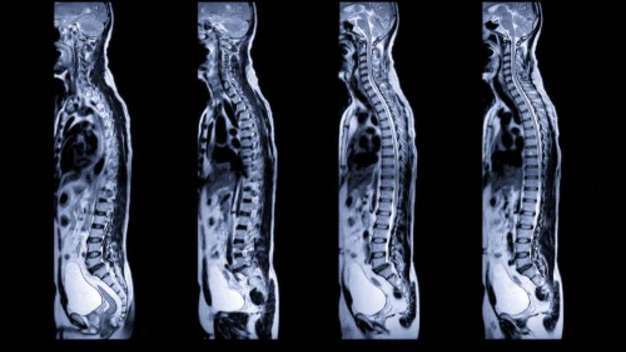
Thoracic Facet Loose Body

The same is true with the spine. There are things that are not seen on an x-ray or even MRI scans. The endoscopic view can reveal things never observed with open or “minimally invasive” surgery. Examples of such things include osteophytes that impinge and hypertrophic capsule and synovium that get pinched in the facet joints.
The video link below shows a loose body inside of a painful thoracic facet joint. The facet block of this joint gave complete relief of the mid-back pain. There is no way to definitively know if the loose body is the underlying cause of pain in this thoracic facet, but it is not supposed to be there and could be the cause of pain. This is an example of a small problem that can cause big pain.
Endoscopic spine surgery is the only way to visualize these small problems that can cause big pain. This is the type of pain problem that doesn’t usually respond to physical therapy, injections, or other conservative measures. The source of pain just remains a mystery until someone is willing to take a look inside of the facet joint, identify the problem, and treat it definitively.
