Could a Discogram be needed?

Are there findings on the physical exam or MRI that might suggest the need for a discogram ?
Yes there are. There are some things that are worth looking for during the physical exam and MRI review that should convince you to do a discogram, especially when you get a proper history.
What is the “proper” history? The proper history is one that describes chronic low back pain that has been present for at least six months, although the duration is usually much longer. The pain is described as deep in the midline of the low back and possibly felt radiating to the “tailbone”. Sitting for any length of time is usually the most uncomfortable position, particularly in a car. This pain can come-and-go for no particular reason and is usually aggravated by physical therapy. Sometimes the pain is accompanied by a sense of “giving away”. The pain can “come and go” for years and most of the time there will be a history of injury or minor trauma that started the back pain.
The physical exam is relatively nonspecific with one exception. When the patient is examined in the prone position, the deep midline pain can be elicited by pushing directly on one of the spinous processes or skin between the spinous processes (interlaminar space). Firm pressure will recreate the deep midline back pain.
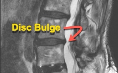
The MRI will most likely be fairly benign in appearance with findings that the radiologist may not even mention. This is one of the reasons that you must personally examine the MRI films and not just read the report. You may see some narrowing of the disc spaces of the lumbar spine and darkening of the disc spaces on the T2 slices that suggest some dehydration, but the MRI finding that correlates best with an annular tear is a disc bulge. Yes, you are correct, bulges are commonly seen on the MRI. However, it is the MRI finding of a bulge taken in context with the proper history and a physical exam in the prone position that indicates further investigation.
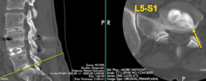
You may be surprised at how many annular tears you will discover with a discogram, like my example below, where the only mention on the radiology report was some darkening of the discs with bulges.
See L5-S1 below.

